A giant ureteric calculus successfully removed by mini-endoscopic combined intrarenal surgery: A case report
Giant ScienceDirect's AI-generated Topic Pages" class="topic-link" style="margin: 0px; padding: 0px; text-decoration-line: underline; text-decoration-thickness: 1px; text-decoration-color: rgb(46, 46, 46); color: rgb(46, 46, 46); word-break: break-word; text-underline-offset: 1px;">ureteric calculi are very extremely rare and typically diagnosed in stones greater than 5 cm in length or 50 g in weight. They may occur as a consequence of tuberculosis, ScienceDirect's AI-generated Topic Pages" class="topic-link" style="margin: 0px; padding: 0px; text-decoration-line: underline; text-decoration-thickness: 1px; text-decoration-color: rgb(46, 46, 46); color: rgb(46, 46, 46); word-break: break-word; text-underline-offset: 1px;">ureteroceles, an ScienceDirect's AI-generated Topic Pages" class="topic-link" style="margin: 0px; padding: 0px; text-decoration-line: underline; text-decoration-thickness: 1px; text-decoration-color: rgb(46, 46, 46); color: rgb(46, 46, 46); word-break: break-word; text-underline-offset: 1px;">ectopic ureter, a benign ScienceDirect's AI-generated Topic Pages" class="topic-link" style="margin: 0px; padding: 0px; text-decoration-line: underline; text-decoration-thickness: 1px; text-decoration-color: rgb(46, 46, 46); color: rgb(46, 46, 46); word-break: break-word; text-underline-offset: 1px;">ureteral polyp, or in the absence of any ScienceDirect's AI-generated Topic Pages" class="topic-link" style="margin: 0px; padding: 0px; text-decoration-line: underline; text-decoration-thickness: 1px; text-decoration-color: rgb(46, 46, 46); color: rgb(46, 46, 46); word-break: break-word; text-underline-offset: 1px;">anatomical abnormalities. Traditional approaches to treating giant ureteral stones are based on the function of the affected kidney and may necessitate either active stone removal or a simple ScienceDirect's AI-generated Topic Pages" class="topic-link" style="margin: 0px; padding: 0px; text-decoration-line: underline; text-decoration-thickness: 1px; text-decoration-color: rgb(46, 46, 46); color: rgb(46, 46, 46); word-break: break-word; text-underline-offset: 1px;">nephroureterectomy.
As a consequence of breakthroughs in urologic interventional techniques and technologies, the ways to eradicate urolithiasis have increased. Miniaturized endoscopic combined intrarenal surgery (mini-ECIRS) is a novel therapeutic option that has become increasingly acceptable for the removal of large or complex ScienceDirect's AI-generated Topic Pages" class="topic-link" style="margin: 0px; padding: 0px; text-decoration-line: underline; text-decoration-thickness: 1px; text-decoration-color: rgb(46, 46, 46); color: rgb(46, 46, 46); word-break: break-word; text-underline-offset: 1px;">nephrolithiasis. To the best of our knowledge, this is the first clinical case reporting mini-ECIRS in a patient with a giant ureteric calculus.
Case report:
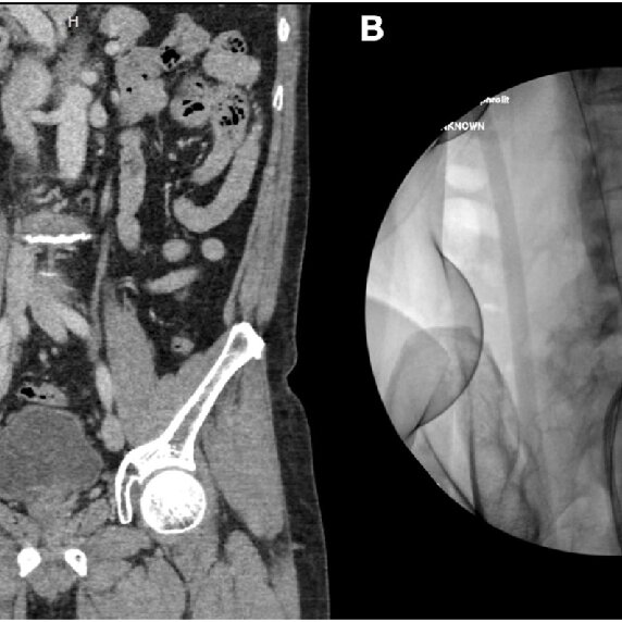
A 58-year-old male was referred to our department with a large ScienceDirect's AI-generated Topic Pages" class="topic-link" style="margin: 0px; padding: 0px; text-decoration-line: underline; text-decoration-thickness: 1px; text-decoration-color: rgb(46, 46, 46); color: rgb(46, 46, 46); word-break: break-word; text-underline-offset: 1px;">ureteral stone that was detected during a routine medical examination. He was in excellent mental and physical health. ScienceDirect's AI-generated Topic Pages" class="topic-link" style="margin: 0px; padding: 0px; text-decoration-line: underline; text-decoration-thickness: 1px; text-decoration-color: rgb(46, 46, 46); color: rgb(46, 46, 46); word-break: break-word; text-underline-offset: 1px;">Urinalysis showed modest ScienceDirect's AI-generated Topic Pages" class="topic-link" style="margin: 0px; padding: 0px; text-decoration-line: underline; text-decoration-thickness: 1px; text-decoration-color: rgb(46, 46, 46); color: rgb(46, 46, 46); word-break: break-word; text-underline-offset: 1px;">pyuria but negative ScienceDirect's AI-generated Topic Pages" class="topic-link" style="margin: 0px; padding: 0px; text-decoration-line: underline; text-decoration-thickness: 1px; text-decoration-color: rgb(46, 46, 46); color: rgb(46, 46, 46); word-break: break-word; text-underline-offset: 1px;">urine cultures. The creatinine level in the blood was 1.1 mg/dL. A ScienceDirect's AI-generated Topic Pages" class="topic-link" style="margin: 0px; padding: 0px; text-decoration-line: underline; text-decoration-thickness: 1px; text-decoration-color: rgb(46, 46, 46); color: rgb(46, 46, 46); word-break: break-word; text-underline-offset: 1px;">computed tomography scan revealed a calculus that was 11 × 12 × 67 mm3 in size with a density of 680 Hounsfield units at the right proximal ScienceDirect's AI-generated Topic Pages" class="topic-link" style="margin: 0px; padding: 0px; text-decoration-line: underline; text-decoration-thickness: 1px; text-decoration-color: rgb(46, 46, 46); color: rgb(46, 46, 46); word-break: break-word; text-underline-offset: 1px;">ureter as shown in9fig 1). Excretion was mildly decreased in the right kidney, but normal excretion was observed on the left side. After all, discussing the various treatment options with the patient, we concluded together that mini-ECIRS would be preferred.

Intravenous prophylactic antibiotics (cefuroxime) were administered during the induction period of ScienceDirect's AI-generated Topic Pages" class="topic-link" style="margin: 0px; padding: 0px; text-decoration-line: underline; text-decoration-thickness: 1px; text-decoration-color: rgb(46, 46, 46); color: rgb(46, 46, 46); word-break: break-word; text-underline-offset: 1px;">general anesthesia. The patient was placed in the Galdakao-modified supine Valdivia position, in which the right flank was raised, and the buttock and leg were set in an asymmetrical ScienceDirect's AI-generated Topic Pages" class="topic-link" style="margin: 0px; padding: 0px; text-decoration-line: underline; text-decoration-thickness: 1px; text-decoration-color: rgb(46, 46, 46); color: rgb(46, 46, 46); word-break: break-word; text-underline-offset: 1px;">lithotomy position. Two operating instruments were used simultaneously by two urologists: one used a miniature nephroscope, while the other used a flexible ureteroscope (fURS).
First, a ScienceDirect's AI-generated Topic Pages" class="topic-link" style="margin: 0px; padding: 0px; text-decoration-line: underline; text-decoration-thickness: 1px; text-decoration-color: rgb(46, 46, 46); color: rgb(46, 46, 46); word-break: break-word; text-underline-offset: 1px;">cystoscopy was carried out to gain retrograde access, and a retrograde ScienceDirect's AI-generated Topic Pages" class="topic-link" style="margin: 0px; padding: 0px; text-decoration-line: underline; text-decoration-thickness: 1px; text-decoration-color: rgb(46, 46, 46); color: rgb(46, 46, 46); word-break: break-word; text-underline-offset: 1px;">pyelogram was performed to delineate the ureter and pelvicalyceal system. for, Two-hybrid guidewires (Sensor™, Boston Scientific, Natick, MA) were then inserted into the ureteric orifice to reach the renal pelvis. A digital fURS, LithoVue™ (Boston Scientific, Marlborough, MA), was introduced over one guidewire until it made contact with the giant calculus. The stone pushed retrogradely into the renal pelvis by advancing the fURS without difficulty. The second urologist performed an ultrasound-guided percutaneous puncture using an 18 G metallic needlepoint to the lower pole of the kidney. Tract dilatation was achieved over the guidewire to establish a 15 F working channel under direct visualization via the fURS. ScienceDirect's AI-generated Topic Pages" class="topic-link" style="margin: 0px; padding: 0px; text-decoration-line: underline; text-decoration-thickness: 1px; text-decoration-color: rgb(46, 46, 46); color: rgb(46, 46, 46); word-break: break-word; text-underline-offset: 1px;">Laser lithotripsy was performed using the fragmentation technique with a 120 W ScienceDirect's AI-generated Topic Pages" class="topic-link" style="margin: 0px; padding: 0px; text-decoration-line: underline; text-decoration-thickness: 1px; text-decoration-color: rgb(46, 46, 46); color: rgb(46, 46, 46); word-break: break-word; text-underline-offset: 1px;">holmium laser (Lumenis, San Jose, CA) and a 550 μm core laser fiber with an energy of 1.5 J and a rate of 30 Hz as shown in fig2. Finally, all the pieces of stone were extracted with graspers through a 12 Fr nephroscope MIP-M system (Karl Storz, City, Germany) or by the Venturi effect. At the end of the procedure, a retrograde 6Fr double-J stent was placed. We obtained plain films of the abdomen to assess residual stones and confirm that the patient kept the double-J stent in the appropriate position (Fig 3). The patient made an uneventful ScienceDirect's AI-generated Topic Pages" class="topic-link" style="margin: 0px; padding: 0px; text-decoration-line: underline; text-decoration-thickness: 1px; text-decoration-color: rgb(46, 46, 46); color: rgb(46, 46, 46); word-break: break-word; text-underline-offset: 1px;">postoperative recovery. The stent was been removed cystoscopically at 6 weeks ScienceDirect's AI-generated Topic Pages" class="topic-link" style="margin: 0px; padding: 0px; text-decoration-line: underline; text-decoration-thickness: 1px; text-decoration-color: rgb(46, 46, 46); color: rgb(46, 46, 46); word-break: break-word; text-underline-offset: 1px;">after discharge.

Fig. 2. Miniature nephoscope view of the giant calculus.

Fig. 3. Post-operative abdominal plain radiography showing no evidence of residual calculi.
Discussion:
- Ureteral calculi are stones made up of crystals in the renal collecting system that then migrate down the ScienceDirect's AI-generated Topic Pages" class="topic-link" style="margin: 0px; padding: 0px; text-decoration-line: underline; text-decoration-thickness: 1px; text-decoration-color: rgb(46, 46, 46); color: rgb(46, 46, 46); word-break: break-word; text-underline-offset: 1px;">ureter.
- They are more likely to become stuck in locations where the ureter is at its narrowest. In general, stones that are less than 4 mm in size are small enough to pass spontaneously in most patients.
- However, for stones greater than 7 mm in width, the probability of spontaneous passage is significantly lower. The best solution for a giant ureteric calculus is controversial.
- The open approach is the preferred choice for most surgeons; however, extracorporeal shock wave ScienceDirect's AI-generated Topic Pages" class="topic-link" style="margin: 0px; padding: 0px; text-decoration-line: underline; text-decoration-thickness: 1px; text-decoration-color: rgb(46, 46, 46); color: rgb(46, 46, 46); word-break: break-word; text-underline-offset: 1px;">lithotripsy, transurethral lithotripsy, ScienceDirect's AI-generated Topic Pages" class="topic-link" style="margin: 0px; padding: 0px; text-decoration-line: underline; text-decoration-thickness: 1px; text-decoration-color: rgb(46, 46, 46); color: rgb(46, 46, 46); word-break: break-word; text-underline-offset: 1px;">percutaneous nephrolithotomy (PCNL), and ScienceDirect's AI-generated Topic Pages" class="topic-link" style="margin: 0px; padding: 0px; text-decoration-line: underline; text-decoration-thickness: 1px; text-decoration-color: rgb(46, 46, 46); color: rgb(46, 46, 46); word-break: break-word; text-underline-offset: 1px;">laparoscopic surgery have also been reported. Notwithstanding, there are no reports of mini-ECIRS for the management of this interesting calculus.
- ECIRS was the first described by Scoffone in 2008 to standardize the synergist procedure between retrograde intrarenal surgery and PCNL for large and/or complex urolithiasis to improve efficiency and safety.
- This combination technique facilitates lithotripsy while keeping the patient in a single position and accessing the collecting system only once.
- The distinguishing benefits of performing the double scopes demonstrated in our case were the relocation of the giant stone upward into the renal pelvis, ureteroscopy-assisted puncture, and the retrieval of any fragments of broken stone that scattered in the different calyx that evades multiple tracts and may cause related bleeding. Moreover, we employed the small access sheath (15 Fr) of a miniature nephroscope, which has significant advantages in terms of reducing the likelihood of kidney injury, bleeding, and the requirement for transfusion.
- The nephrostomy-free or “tubeless” option was considered appropriate in this case as it helps reduce ScienceDirect's AI-generated Topic Pages" class="topic-link" style="margin: 0px; padding: 0px; text-decoration-line: underline; text-decoration-thickness: 1px; text-decoration-color: rgb(46, 46, 46); color: rgb(46, 46, 46); word-break: break-word; text-underline-offset: 1px;">postoperative pain, analgesia requirements, and hospital stay. We confirm the excellent results of the mini-ECIRS procedure by the absence of any pieces of residual stone in plain films of the abdomen administered post-surgery.
- Conclusion
Hence, We successfully treated a giant ureteric calculus using mini-ECIRS. This novel technique is safe and feasible, with low morbidity.
Story Source:
Materials provided by Urology Case Reports. The original text of this story is licensed under a Creative Commons License. Note: Content may be edited for style and length.
Journal Reference: Science direct
0 Comments