New Imaging Technique Provides Breakthrough in Head and Neck Cancer Prognostics

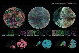
A groundbreaking study by researchers from the University of Helsinki, the University of Turku, and Germany’s Max Planck Institute for Molecular Biomedicine has introduced an advanced imaging technique for analyzing head and neck squamous cell carcinoma. Using machine learning, this method examines cancer and surrounding tissue at the level of individual cells, creating a unique 'fingerprint' for each patient to better predict prognosis and guide treatment.
Innovative Approach to Cancer Analysis
The technology behind this study combines indicators of cancer cell behavior with detailed analyses of tumor structure and surrounding healthy tissue architecture. The study identified two previously undetected patient groups, one with an exceptionally good prognosis and the other with a poor prognosis. The contrast in outcomes was attributed to specific combinations of cancer cell states and the composition of the surrounding tissue. In cases with poor prognosis, the cancer’s aggressiveness was linked to signaling between the cancerous and healthy connective tissue via the epidermal growth factor (EGF).
Implications for Personalized Treatment
“These findings represent a breakthrough in cancer diagnostics, illustrating that specific interactions between malignant and healthy tissue cells have a profound impact on cancer progression,” said Research Director Sara Wickström. “Our method can accurately identify patients with poor prognosis who may benefit from aggressive treatment and, conversely, patients who might manage well with less invasive options, preserving quality of life.”
Path Forward in Diagnostics
The research team is now developing a diagnostic test based on this technology for use in clinical settings. They are also exploring its applicability to other cancers, such as colorectal cancer, supported by funding from Business Finland’s Research to Business program. Wickström noted, “This technology leverages machine learning and spatial biology to process extensive data on patient samples and cells, promising a transformative impact on cancer diagnostics and treatment strategies.” She emphasized that the method is cost-effective, relying on readily available antibody staining and a proprietary algorithm.
Story Source:
Materials provided by University of Helsinki. The original text of this story is licensed under a Creative Commons License. Note: Content may be edited for style and length.
Journal Reference:
- Karolina Punovuori, Fabien Bertillot, Yekaterina A. Miroshnikova, Mirjam I. Binner, Satu-Marja Myllymäki, Gautier Follain, Kai Kruse, Johannes Routila, Teemu Huusko, Teijo Pellinen, Jaana Hagström, Noemi Kedei, Sami Ventelä, Antti Mäkitie, Johanna Ivaska, Sara A. Wickström. Multiparameter imaging reveals clinically relevant cancer cell-stroma interaction dynamics in head and neck cancer. Cell, 2024; DOI: 10.1016/j.cell.2024.09.046

0 Comments